
Two papers (this and this) and an editorial in the latest issue of Archives of Internal Medicine examine the cancer risks associated with the use of computed tomography (CT) examinations.1
Ionizing radiation increases the risk for developing cancer. There is direct evidence from atomic bomb survivors in Japan in 1945 and from nuclear accidents such as the one in Chernobyl in 1986. There are no studies directly demonstrating an increased cancer risk from the diagnostic use of X-rays (either conventional radiographs or computed tomography), as this would require long-term follow-up of a very large number of people. This risk can only be estimated, based on the assumption that there is no lower radiation threshold dose for cancer risk and that a linear correlation exists between dose and cancer risk. Similarly, we know very little about the actual radiation doses that patients receive during a diagnostic CT scan.
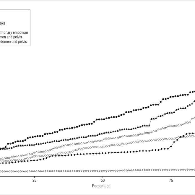
The paper by Rebecca Smith-Bindman and her coworkers looked at the radiation doses associated with the 11 most common computed tomography examinations in four hospitals in the San Francisco Bay Area. Radiation doses to individual patients were not measured directly, but were estimated using the commonly used “effective dose”. Median effective doses for routine computed tomography of the head, chest and abdomen-pelvis were 2, 8 and 16 mSv. The highest median effective doses (31 mSv) were used for multiphase CTs of the abdomen-pelvis (range 6-90 mSv). The 8 mSv dose for a routine chest CT is equivalent to 119 conventional chest X-rays. Interestingly, there was wide variation of effective doses used both between hospitals and within the same institution (with a mean 13-fold variation between the highest and lowest dose for each CT study type).
Based on these doses, the authors then estimated the increased risk for radiation-induced cancer following a CT examination. This risk is higher in younger people and in women. Women have a higher risk to develop lung cancer following radiation exposure and they have the added risk of developing breast cancer. The authors estimated that it would for example require approximately 620 CT scans for ruling out pulmonary embolism in 40-year-old women to induce one cancer.
The second paper by Amy Berrington de Gonzalez and coworkers tried to calculate the total number of CT scans performed in the US in 2007 and estimated the increased risk for cancer related to these CT scans based on examination type, age, and sex. They used a similar model to calculate the cancer risk as the first paper. Overall, about 72 million CT scans were performed in the US in 2007. The authors estimated that approximately 29.000 additional cancers will eventually develop, equivalent to approximately 2% of all cancers diagnosed annually in the US.
CT scans have dramatically improved patient care and the conclusions of these two papers should not preclude their use in patients where the potential benefits clearly outweigh the risks mentioned above. But in order to reduce the cancer risk associated with CT scans, the authors of the two papers and the editorial suggest optimizing and standardizing the CT scan procedure (remember the 13-fold difference in dose for the same procedure) and decreasing the number of unnecessary examinations. The latter is most relevant when CT scans are performed as screening procedures.
More blog posts discussing these papers can be found on Nature Blogs and Dr. Len's Cancer Blog.
1 All three papers are available as full-text without a subscription. And none of the papers displays its DOI, which makes linking to them unnecessarily difficult.
References
Smith-Bindman R. Radiation Dose Associated With Common Computed Tomography Examinations and the Associated Lifetime Attributable Risk of Cancer. Arch Intern Med. 2009;169(22):2078. doi:10.1001/archinternmed.2009.427
Berrington De González A. Projected Cancer Risks From Computed Tomographic Scans Performed in the United States in 2007. Arch Intern Med. 2009;169(22):2071. doi:10.1001/archinternmed.2009.440
Redberg RF. Cancer Risks and Radiation Exposure From Computed Tomographic Scans: How Can We Be Sure That the Benefits Outweigh the Risks? Arch Intern Med. 2009;169(22):2049. doi:10.1001/archinternmed.2009.453
